Epidermoid Cyst (Sebaceous Cyst)
Epidermoid cysts, sometimes called sebaceous cysts, contain a soft, “cheesy” material composed of keratin, a protein component of skin, hair, and nails.
Epidermoid cysts form when the top layer of skin (the epidermis) grows into the middle layer of the skin (the dermis). This may occur due to injury or blocked hair follicles.
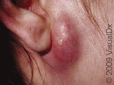
An epidermoid cyst may have no pain or other symptoms associated with it, but if it becomes inflamed or infected, it may grow, become painful and red, and may rupture.
Who's At Risk?
Epidermoid cysts are common and can affect people of any race / ethnicity, age, and sex. They are most common in males, though, and they are uncommon in children.
Signs & Symptoms
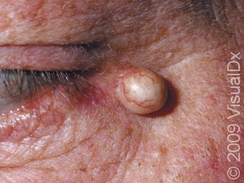
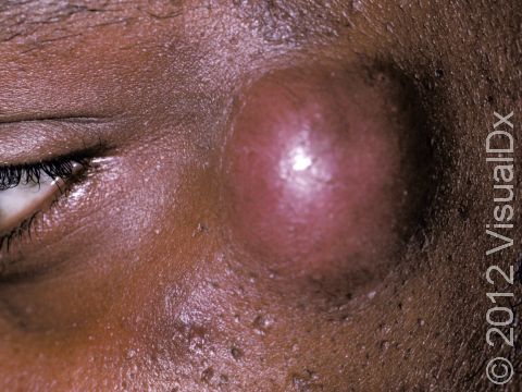
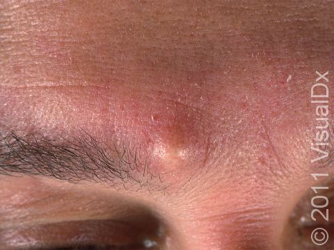
Epidermoid cysts can be located almost anywhere on the body, but they are most common on the face, neck, scalp, and trunk.
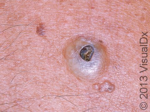
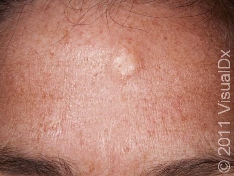
The cyst appears as a dome-shaped, skin-colored nodule (a solid bump) that usually moves when touched and pressed upon. It may have an opening in the center that looks like an enlarged pore.
If the cyst is picked at or bumped or if it is infected, it can become red and may be tender.
Self-Care Guidelines
No self-care is necessary. It is important, though, to avoid picking at or popping the cyst because further inflammation and even infection may result.
Treatments
Your medical professional may:
- Inject the cyst with steroids to reduce inflammation.
- Drain the cyst to decrease its size, but this is a temporary measure. Eventually, the cyst will refill with the cheesy contents because the lining of the cyst has not been removed.
- Remove (excise) the cyst surgically.
- Prescribe antibiotics if the cyst is infected.
Visit Urgency
See your medical professional if you have a cyst that becomes inflamed or painful. Also see a medical professional if you develop multiple epidermoid cysts, especially if they run in your family.
Trusted Links
References
Bolognia J, Schaffer JV, Cerroni L. Dermatology. 4th ed. Philadelphia, PA: Elsevier; 2018.
James WD, Elston D, Treat JR, Rosenbach MA. Andrew’s Diseases of the Skin. 13th ed. Philadelphia, PA: Elsevier; 2019.
Kang S, Amagai M, Bruckner AL, et al. Fitzpatrick’s Dermatology. 9th ed. New York, NY: McGraw-Hill Education; 2019.
Last modified on June 26th, 2024 at 1:08 pm
Not sure what to look for?
Try our new Rash and Skin Condition Finder
