Managing Fungal Skin Infections: Tips for Prevention and Treatment
Fungal infections of the skin are a common complaint among patients visiting a dermatologist. In this article, we will discuss two of the most common types of fungal skin infections: tinea skin infections and pityriasis versicolor. We will also discuss some of the tests and treatments that dermatologists can use for each condition.
Tinea Skin Infections (Dermatophytes)
Tinea skin infections are caused by a group of fungi known as dermatophytes, which include Trichophyton, Epidermophyton, and Microsporum. These fungi can infest the top layer of the skin, feeding on dead skin cells, and spread out centrifugally over time. Depending on the location the fungus colonizes, dermatophyte skin infections have different names.
Tinea pedis refers to a dermatophyte infection on the foot. There are several known patterns, such as interdigital (between the toes) or “moccasin” (on the sole and/or sides of the foot). The common name for tinea pedis is athlete’s foot.
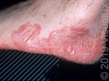
(“Moccasin” distribution of tinea pedis)
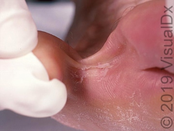
(Interdigital distribution of tinea pedis)
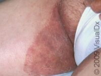
Tinea cruris refers to a dermatophyte infection of groin and is commonly referred to as jock itch.
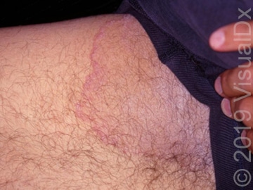
Tinea capitis refers to a dermatophyte infection of the scalp. This type of infection is less common in adults and is more difficult to treat.

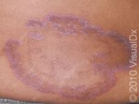
Tinea corporis refers to a dermatophyte infection found on the trunk or extremities. This is also known as ringworm.
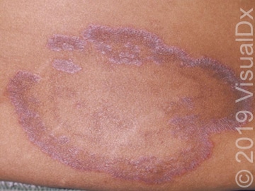
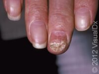
Onychomycosis is a fungal infection of the toenail. Like tinea capitis, it can be more challenging to treat than other dermatophyte infections.
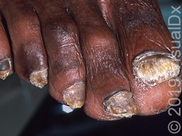
All of the dermatophyte infections can be treated with antifungal medications such as terbinafine, but the route of administration can vary. Typically, these antifungals are used topically; however, tinea capitis and onychomycosis are particularly challenging to treat and typically require oral antifungal medications in order to eradicate the fungus.
Pityriasis Versicolor
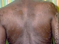
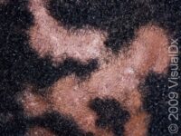
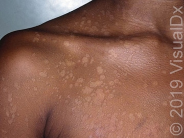
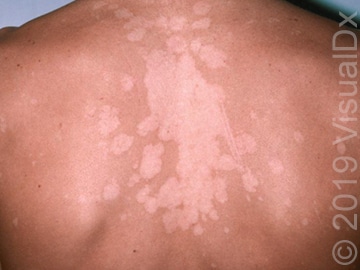
The other broad category of fungus is pityriasis versicolor. This infection is caused by the species Malassezia furfur and has a different presentation and treatment. Pityriasis versicolor usually presents as hyper- or hypopigmented, circular, scaly lesions, as opposed to the red or pink lesions caused by dermatophyte skin infections. The treatment for pityriasis versicolor includes antifungal shampoos with ingredients such as selenium sulfide or pyrithione zinc that can kill the fungus that causes this infection.
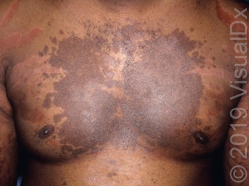
Testing for Tinea or Pityriasis Versicolor
If your dermatologist suspects that you might have a fungal skin infections, he/she might do a potassium hydroxide (KOH) scrape test. The doctor will use a small scalpel blade to scrape off some of the scale from the lesion onto a glass slide. KOH is then applied to the debris, which dissolves the skin cells and leaves behind just the fungus, if any is present. The dermatologist will then look at the slide under the microscope to check for patterns identifying the type of fungal infection present. These tests aren’t always 100% accurate, and the diagnostic accuracy can vary based on the skill of the dermatologist doing the test.
Last modified on August 31st, 2023 at 4:02 pm
