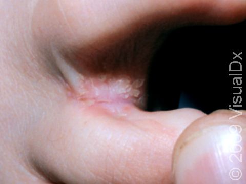
This image displays scaling between the toes typical of tinea pedis (athlete's foot).
Graphic content
Please click to view.
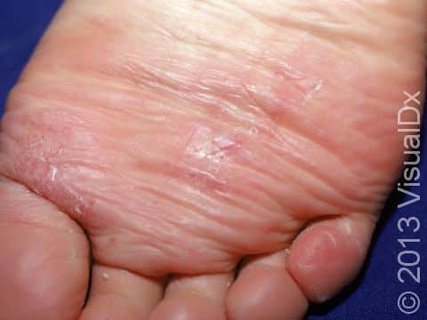
This image displays scaly, slightly elevated lesions typical of tinea pedis (athlete's foot).
The space between the 4th and 5th toe is a frequent location of the start of athlete's foot (tinea pedis).
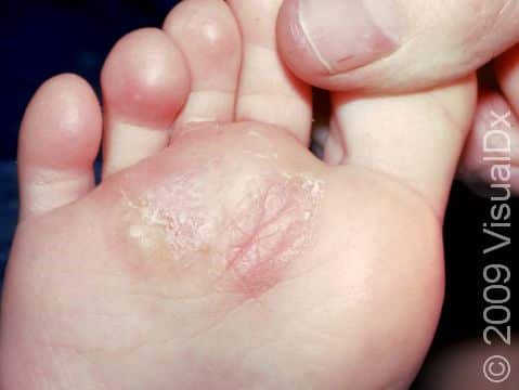
This image displays tinea (athlete's foot) on the bottom area of the foot creeping toward the space between the second and third toes.
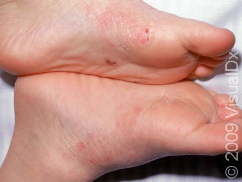
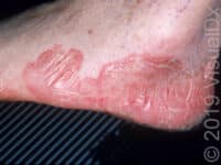
This image displays red, scaly patches on the instep soles typical of tinea pedis (athlete's foot).
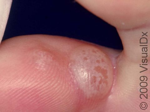
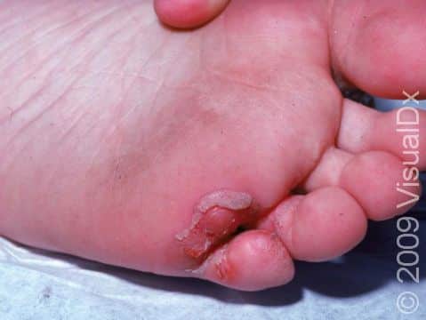
Athlete's foot (tinea pedis) can cause blisters, such as this case between the toes.
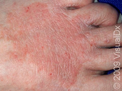
The circular shape of this red, scaling area of skin on the back of the foot demonstrates why tinea is often called "ringworm."
Graphic content
Athlete's Foot (Tinea Pedis)
Athlete’s foot (tinea pedis), also known as ringworm of the foot, is a superficial fungal infection of the skin of the foot. Athlete’s foot is the most common fungal disease in humans, although it is rare in young children.
Athlete’s foot may be passed to humans by direct contact with infected people, infected animals, contaminated objects (such as towels or locker room floors), or the soil.
Who's At Risk?
Athlete’s foot may occur in people of all ages, races / ethnicities, and sexes. Young children rarely develop athlete’s foot, but it is frequently seen in teens and adults. In addition, athlete’s foot is more common in males than in females.
Some conditions make athlete’s foot infections more likely to occur:
- Living in warm, humid climates
- Using public or community pools or showers
- Wearing tight, nonventilated footwear
- Sweating profusely
- Having diabetes or a weakened immune system
Signs & Symptoms
The most common locations for athlete’s foot include the:
- Spaces (webs) between the toes.
- Soles of the feet.
- Tops of the feet (very unusual in children).
Athlete’s foot may affect one or both feet. It can look different, depending on which part of the foot (or feet) is involved and which fungus caused the infection:
- Between the toes (the interdigital spaces), athlete’s foot may appear as inflamed, scaly, and soggy tissue. Splitting of the skin, called fissures, may be present between or under the toes. This form of athlete’s foot tends to be quite itchy.
- On the sole of the foot (the plantar surface), athlete’s foot may appear as skin color changes with scales ranging from mild to widespread (diffuse). In lighter skin colors, the border of the affected area is often pink or red. In darker skin colors, it may be dark red, purple, grayish, or dark brown.
- On the top of the foot, athlete’s foot appears as one or more scaly patches with skin color changes ranging in size from 1-5 cm. The border of the affected skin may be raised and may contain bumps, blisters, or scabs. Often, the central portion of the lesion is clear, leading to a ring-like shape and the descriptive (but inaccurate) name “ringworm.”
- Another type of tinea infection, called bullous tinea pedis, appears as painful and itchy blisters on the arch and/or the ball of the foot.
- The most severe form of the infection, called ulcerative tinea pedis, appears as painful blisters, pus-filled bumps (pustules), and shallow open sores (ulcers). These lesions are especially common between the toes but may involve the entire sole. Because of the numerous breaks in the skin, lesions commonly become infected with bacteria. Ulcerative tinea pedis occurs most frequently in people with diabetes and others with weakened immune systems.
Self-Care Guidelines
If you suspect that your child has athlete’s foot, you might try one of the following over-the-counter antifungal creams:
- Terbinafine (eg, Lamisil)
- Clotrimazole (eg, Lotrimin AF)
- Miconazole (eg, Desenex, Micatin)
Apply the cream between the toes and to the soles of both feet for at least 2 weeks after the areas are completely clear of lesions.
In addition, try to keep your child’s feet dry, creating conditions where the fungus cannot live and grow. Have your child try the following:
- Wash their feet daily and dry them well.
- Use a separate towel for the feet than the rest of the body, and do not share this towel with anyone else.
- Wear cotton socks and change them once or twice a day, or even more often if they become damp.
- Avoid wearing shoes made of synthetic materials such as rubber or vinyl, and consider throwing out old footwear or treating shoes with antifungal powder.
- Wear sandals when possible.
- Wear protective footwear in locker rooms and public or community pools and showers.
Treatments
To confirm the diagnosis of athlete’s foot, your child’s medical professional may scrape some surface skin material (scales) onto a glass slide and examine them under a microscope. This procedure, called a potassium hydroxide (KOH) preparation, allows them to look for signs of fungal infection.
Once the diagnosis of athlete’s foot has been confirmed, the medical professional will likely prescribe treatment with an antifungal medication such as:
- Econazole (Spectazole).
- Oxiconazole (Oxistat).
- Ciclopirox (eg, Loprox).
- Ketoconazole (eg, Nizoral).
- Naftifine (Naftin).
- Butenafine (Lotrimin, Mentax).
- Luliconazole (Luzu).
- Sertaconazole (Ertaczo).
- Sulconazole (Exelderm).
Other topical medications your child’s medical professional may consider include:
- Compounds containing salicylic acid or urea to help dissolve the scale and allow the antifungal cream to penetrate better into the skin.
- Solutions containing aluminum chloride, which reduce sweating of the foot.
- Antibiotic creams to prevent or treat bacterial infections, if present.
Rarely, more extensive infections or those not improving with topical antifungal medications may require 3-4 weeks of treatment with oral antifungal pills, including:
- Terbinafine (Lamisil; in children 4 years and older).
- Itraconazole (Sporanox).
The infection should go away within 4-6 weeks after using effective treatment.
Visit Urgency
If the lesions do not improve after 2 weeks of applying over-the-counter antifungal cream or if they are exceptionally itchy or painful, see your child’s medical professional for an evaluation. If your child has blisters, pustules, and/or ulcers on the feet, see their medical professional as soon as possible.
Trusted Links
References
Bolognia J, Schaffer JV, Cerroni L. Dermatology. 4th ed. Philadelphia, PA: Elsevier; 2018.
James WD, Elston D, Treat JR, Rosenbach MA. Andrew’s Diseases of the Skin. 13th ed. Philadelphia, PA: Elsevier; 2019.
Kang S, Amagai M, Bruckner AL, et al. Fitzpatrick’s Dermatology. 9th ed. New York, NY: McGraw-Hill Education; 2019.
Paller A, Mancini A. Paller and Mancini: Hurwitz Clinical Pediatric Dermatology. 6th ed. St. Louis, MO: Elsevier; 2022.
Last modified on June 14th, 2024 at 1:13 pm
Related Articles
Not sure what to look for?
Try our new Rash and Skin Condition Finder