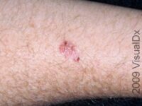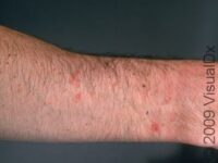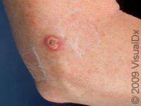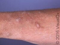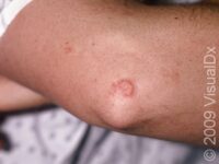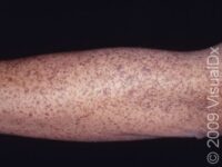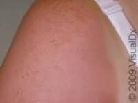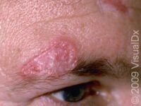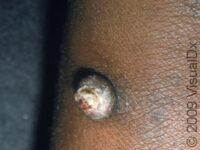
Actinic Keratosis (Solar Keratosis)
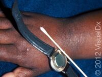
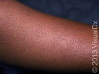
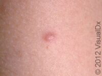
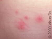
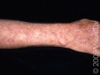
The forearm is a very common area for sun damage and actinic keratoses.
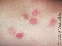
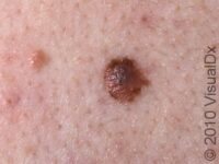
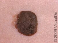
VisualDx has identified the following matches. Tap each one to see more images for more information.
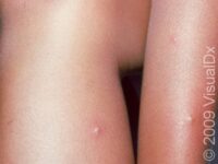
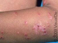
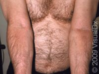
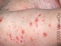
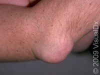
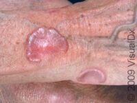
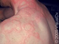
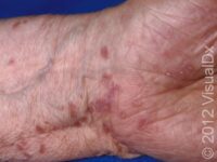
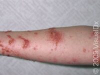
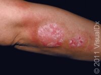
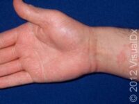
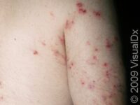
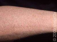
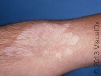
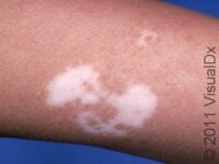
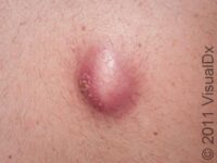
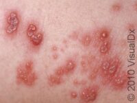
On the page below, you will find links to information on the conditions that can affect the arm in adults. Like other large parts of the skin, sun damage causes conditions ranging from sunburn to skin cancer. Many chronic conditions can affect the arm. Psoriasis will cause large raised patches, usually around the elbow. Ichthyosis vulgaris is a hereditary condition that causes the skin to appear dry and scaly, sometimes like the scales of a fish. Vitiligo involves the destruction of pigmented cells (melanocytes) in the skin. It is more pronounced in darker-skinned people and typically takes a patchy glove-like pattern on the arms. Keratosis pilaris is often seen on the arms and has the appearance of goose pimples that do not go away.