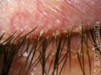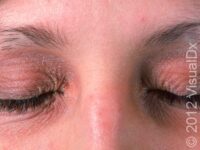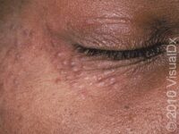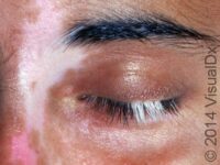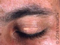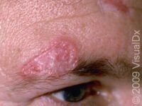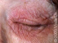
Allergic Contact Dermatitis
The thin eyelid skin is a frequent site for allergic contact dermatitis due to inadvertent touching the eyelids…
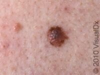
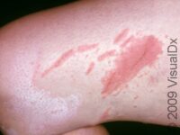
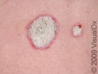
VisualDx has identified the following matches. Tap each one to see more images for more information.
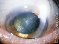
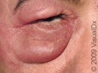
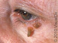
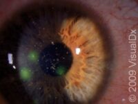
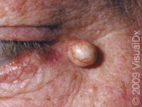
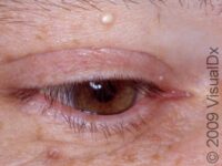
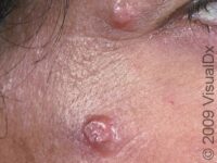
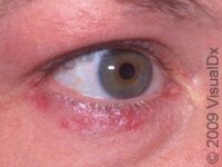
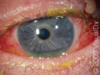
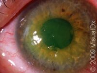
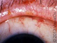
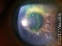
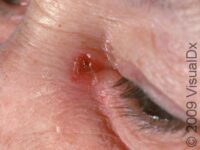
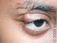
Click the pictures or links below for information about conditions that can affect the eye in adults. There is an immense variety of potential problems involving the eye. Pink eye (conjunctivitis) is a common and contagious infection that can be caused by a virus or bacteria. The cornea is the clear part of the eye that covers the pupil. It is easily damaged resulting in a corneal abrasion or corneal erosion. The eyelids are rich with lubrication glands. Obstruction of these glands can lead to 2 types of styes, known as a hordeolum or chalazion. In general, it is advisable to see a physician for any sudden change in vision or onset of pain in the eye.